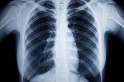
What is an X-Ray?
X-rays are a form of electromagnetic radiation, just like visible light. In a healthcare setting, a machine sends individual x-ray particles called photons. These particles pass through the body. A computer or film is used to record the images created.
Dense structures (such as bone) will block most x-ray particles and appear white. Metal and contrast media (special dye used to highlight areas of the body) will also appear white. Structures containing air will be black, and muscle, fat, and fluid will appear shades of gray.
How is an X-Ray performed?
The test is performed in a hospital radiology department or in the health care provider’s office by an x-ray technologist. The positioning of the patient, x-ray machine, and film depends on the type of study and area of interest. Multiple individual views may be requested.
Much like conventional photography, motion causes blurry images on radiographs; thus, patients may be asked to hold their breath or not move during brief exposure (about 1 second).
When is an X-ray performed?
Before a doctor determines the correct procedure, they must first check your bone images. This will determine the severity of your problem. X-rays are not only for the bones; these are the standard procedures done in some parts of the body:
Head Bones (including the teeth)
- Dental checkup. Dentists use X-rays to inspect for cavities in your teeth. Sometimes, they also use X-rays before preparing for an orthodontics procedure (braces).
- Arthritis. Joint X-rays can reveal proof of arthritis. Regular X-rays can help your doctor discern if your arthritis is worsening.
- Fractures and infections. In most circumstances, infections in bones and teeth and fractures and show up on X-rays.
- Osteoporosis. Special types of X-ray tests can calculate your bone density.
- Bone cancer. X-rays can uncover bone tumors.
Chest
- Lung infections or conditions. Proof of tuberculosis, pneumonia, or lung cancer can appear on chest X-rays.
- Mammography. A special type of X-ray test to examine breast tissue for Breast cancer.
- Enlarged heart. A chest X-ray can find congestive heart failure clearly.
- Blocked blood vessels. Injecting a contrast material** with iodine can help emphasize sections of your circulatory system to examine blocked vessels.
Abdomen
- Digestive tract problems. Injecting a contrast medium** with barium delivered in a drink or an enema can help uncover problems in your digestive system.
- Swallowed items. Xray can locate swallowed objects such as a key or a coin.
** What is contrast material?
In some special cases, this liquid called contrast medium will be administered to you. Contrast mediums, such as barium and iodine, aid outline a specific part of your body on the image output. You may consume the contrast medium or be injected or an enema. An X-ray machine takes images of the colon during a barium enema exam, while an enema tube inserted in the rectum supplies liquid barium.
If abdominal studies are intended, and you have had a barium contrast study (such as a barium enema, upper GI series, or barium swallow) or taken prescriptions containing bismuth (such as Pepto-Bismol) in the last 4 days. In that case, the test may be postponed until the contrast has fully expired.
How to prepare for an X-Ray?
In general, you undress the part that needs examination. Depending on which area is being X-rayed, the technician may ask you to wear a gown during the exam.
Inform the health care provider before the exam if you are pregnant, may be pregnant, or have an IUD inserted.
You will remove all jewelry and wear a hospital gown during the x-ray examination because metal and specific clothing can obscure the images and require repeat studies.
How will the X-Ray feel?
There is no discomfort from x-ray exposure. Patients may be asked to stay in awkward positions for a short time. But for very rare few, exposure to x-rays may cause side effects.
What are some risks of an X-Ray?
Some worry that X-rays aren’t safe because radiation exposure causes cell mutation that leads to cancer. The quantity of radiation exposure during an X-ray depends on the tissue or organ being scanned. Sensitivity to radiation depends on age, with children being more sensitive than adults.
Generally, radiation exposure is low, and the benefits from these tests far outweigh the risks.
For most conventional x-rays, the risk of cancer or defects due to damaged ovarian or sperm cells is superficial. Most professionals feel this low risk is vastly outweighed by the advantages of appropriate imaging.
For some, the injection of a contrast medium may cause side effects such as:
- Warm flushes (feeling of warmth)
- Feeling of lightheadedness
- A metallic taste
- Nausea (feeling of wanting to vomit)
- Hives
- Itching
Very rarely, severe reactions to a contrast medium occur, including:
- Anaphylactic shock
- Cardiac arrest
- Severe low blood pressure
These side effects may not be common to regular people. That is why it is essential to seek medical advice from a doctor or a prescription before an X-ray procedure.
X-rays are checked and regulated for patients to have a minimum amount of radiation exposure needed to get the image. Young children and fetuses are more sensitive to the risks of x-rays. Women should tell healthcare providers if they think they are pregnant.
After the X-ray
You can generally continue normal activities. Regular X-rays usually bear no side effects. If you’re injected with a contrast medium before your X-rays, consume plenty of liquids to eliminate the medium from your body. Contact your doctor if you have discomfort, redness, or swelling at the injection site.
Results
X-rays are mostly digitally saved, which can be viewed on-screen within a few minutes. A radiologist typically views and analyzes the results and sends the report to your doctor. Your medical practitioner will explain the results to you. In emergency cases, your X-ray results can be available to your doctor in minutes.


